
Baltic Vein Clinic offers diagnosis for preventive purposes and in case of suspected disease or to understand the correct method of treatment.
| Different vein diseases can have similar or even identical symptoms. Therefore, vein diagnosis is just as important as treatment. As the largest vein clinic in the sector, the Baltic Vein Clinic offers all modern diagnostic methods to ensure that the selected therapy is accurate, error-free, and as effective as possible. |
If relatives have a history of vein diseases
If only one parent has varicose veins, boys may inherit it in ca. 25% and girls in 62% of cases. If both parents (approx. 90% of cases) suffer from varicose veins, there is a risk of inheriting the disease. Varicose veins may usually skin a generation.
Around the age of 30–35 years
Varicose veins are very common in women during this period (leg vein problems are much less common in men than in women). However, if the disease is inherited, it can progress in girls from the age of 13-14 years.
If you smoke and take contraception
If smokers taking hormonal contraception have varicose veins, the risk of thrombosis increases tenfold. Hormonal contraception is not recommended even for passive smokers. If a woman is prone to varicose veins, hormones will increase the risk. The progesterone contained in the hormonal medicines relaxes the smooth muscle layer in the vein wall. It is more difficult for a vein to ensure correct blood flow from the foot to the body. The reverse blood flow caused by gravitational forces (from top to bottom) starts. It is advisable to have your veins diagnosed before taking hormonal medicines, as the first signs of varicose veins cannot be seen with the naked eye.
After childbirth
Pregnancy is also a risk factor. During this period, hormonal changes occur in the woman's body, and the level of female hormones in the blood rises, as a result, the walls of the veins become weaker. In late pregnancy, when the baby compresses the pelvic veins, additional pressure builds up, which interferes with normal blood circulation, so varicose veins can continue to develop after the baby is born.
If the popliteal vein is compressed for a long time
If you squat for several hours, for example, in a flowerbed, blood circulation is impaired. Athletes who often strain their leg muscles are more likely to have vein problems.
If you are overweight or do manual work
Overweight increases the strain on your legs. Prolonged and regular (more than 4 hours) exposure to heat or physically demanding work can worsen the condition of the veins, as the blood vessels dilate, and their functional abilities decrease.
Wearing high-heeled shoes
High heels contribute not only to flat feet and increased foot sweating (the sweat glands on the balls of the feet near the toes are compressed), but also to varicose veins. Wearing high-heeled shoes strains the calf muscle, which is the main venous pump, and blood congestion is formed in the veins, which promotes the development of varicose veins. It is no accident that women develop varicose veins 3 to 4 times more often.
In case of discomfort
If there is non-specific discomfort in the legs, they feel tired all the time, as if they are filled with lead, there is swelling, aching pain in the calf area (sometimes cramps in the calf muscles).
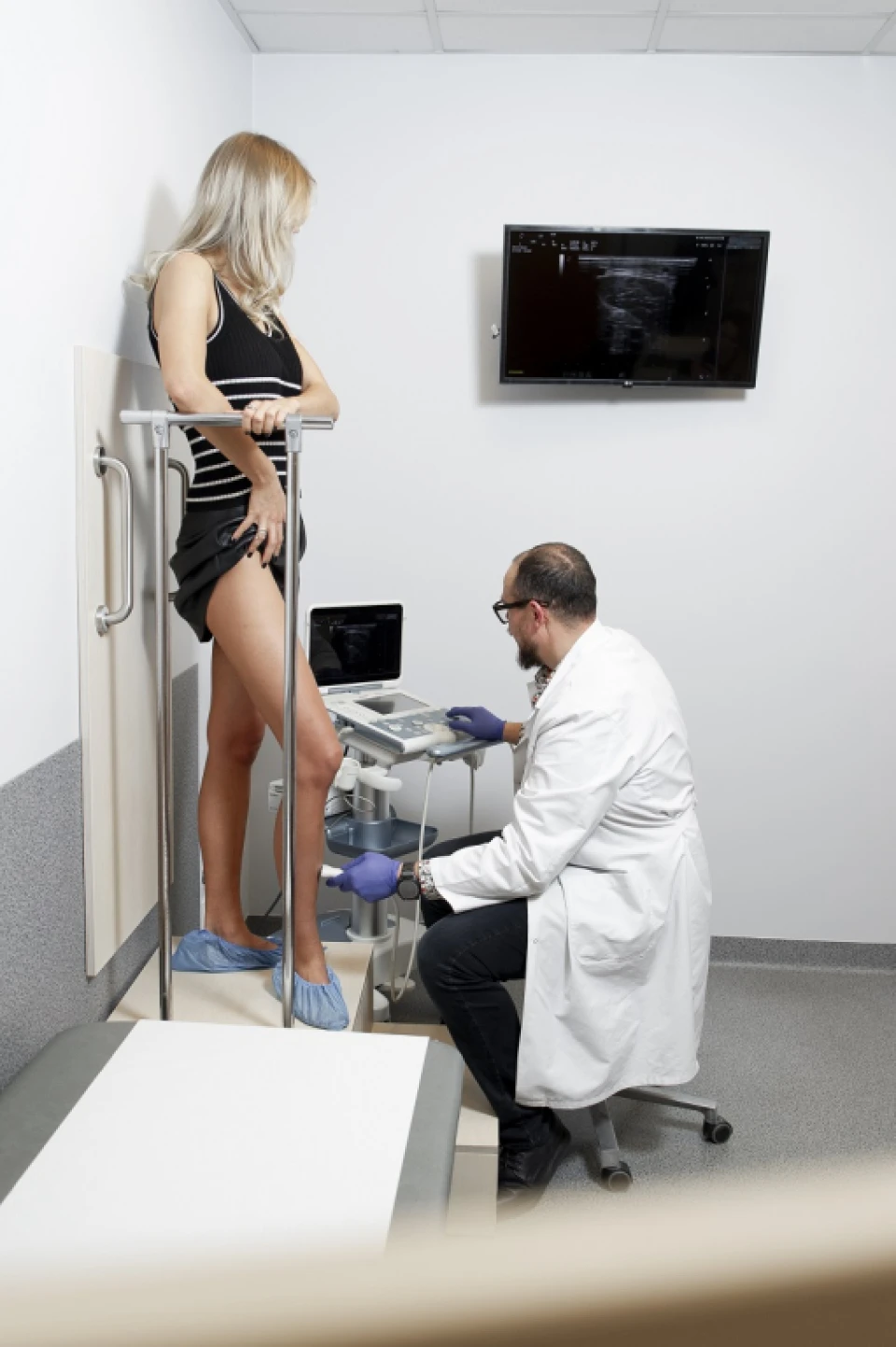
This device is used to measure the blood backflow in the veins and to identify the severity of damage to vein valves and damaged areas of the veins. It allows accurate and prompt assessment of venous reflux in deep and superficial veins, evaluating them visually and diagnosing thrombosis and other vein pathologies. When veins are examined using duplex ultrasound, accurate information about the condition of deep and superficial veins can be obtained within approximately 10-20 minutes.
whether the veins are sick and should be operated;
whether the blood vessels are healthy, located close to the skin and pose a cosmetic issue only.
Bluish veins (spider veins) are sometimes visible in the legs, but they are completely healthy. Most often, thin skin is to blame for this cosmetic defect, as the vascular network is clearly visible through it. In these cases, the appearance is often improved if the skin is lightly tanned.
The duplex ultrasound method is also applied to identify the extent of the planned surgery and to carry out accurate preoperative vein marking. The vein diagnosis only provides accurate information when the patient is examined standing up or in a tilted position with the legs down, so that the force of gravity has a proper effect on the blood flow. The examination report is best represented visually, with marked damaged vein segments on the diagram and indicating both the duration of venous reflux and the vein diameter.
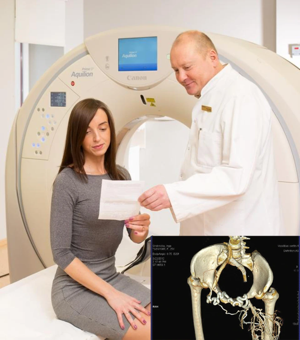
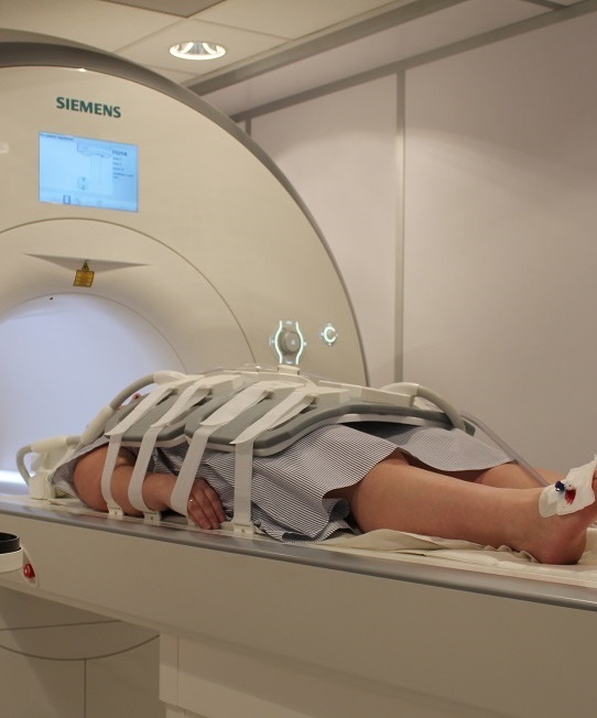
Veselības centrs 4 offers computed tomography to its patients. Computed tomography phlebography is mostly indicated for deep vein thrombosis of unclear aetiology or in case of congenital vein diseases.
Other examination methods allow only to examine the deep veins segmentally, but computed tomography phlebography shows the entire deep vein system.
During the examination, a contrast medium is slowly injected into the superficial vein of the leg with a tourniquet applied so that the contrast medium fills the deep veins. When the appropriate filling has been achieved, as established by CT monitoring, a quick scan of the limb is performed and the enhancement in the deep veins is obtained.
The examination is performed in the computed tomography office of Veselības centrs 4 using a new Siemens Somatom Definition AS computed tomography device.
Angiography is a high-precision medical examination used to evaluate blood vessels—arteries and veins—in detail. It helps determine whether there are narrowings, blockages, blood clots, aneurysms, or other changes that may affect blood circulation. This test is used when a particularly precise assessment of vascular anatomy and blood flow is required.
when deep vein obstruction is suspected;
when highly detailed vascular mapping is necessary;
when a specific vascular surgical or endovascular intervention is planned.
During angiography, a special contrast agent is injected into a blood vessel, and imaging is performed—most commonly using X-ray, computed tomography (CT angiography), or magnetic resonance imaging (MR angiography). The contrast agent makes the blood vessels clearly visible on the images.
The type of examination is selected by the physician based on the patient’s health condition and the diagnostic objective.
Today, angiography is a safe diagnostic method. As with any medical procedure, there are certain risks (such as an allergic reaction to the contrast agent), but these are relatively rare.
Before the examination, the doctor evaluates kidney function, allergy risks, chronic diseases, and any medications being taken.
SIA "Veselības centrs 4" is one of the largest private multi-profile outpatient medical companies in Latvia with 30 years of experience and technologically advanced equipment. The main areas of operation include diverse diagnostics, full-spectrum treatment, modern rehabilitation, and a new concept of preventive and aesthetic medicine.
